BPC-157, TB-500, and GHK-Cu mostly rely on animal and lab work
What do those studies tell you? You see how repair changed. They can't prove what the three peptides do in your body.
BPC-157 Mechanism of Action: Small vessels grew in rats
How might BPC-157 help hurt tissue? It may help small blood vessels grow.
It may also help tendon repair cells multiply [1][2][3]. Those changes could bring blood and repair cells to an injury.
One part of the finding involved nitric oxide. Your body uses it to relax blood vessels.
One study used human blood-vessel cells and rat legs [1]. The cells made more of the parts that help small vessels grow.
The rat legs grew more small vessels. Blood flow returned faster too.
Another study used the rat's main artery [2]. The artery relaxed more as researchers added more BPC-157.
The artery's inner lining released more nitric oxide. That helped the artery open.
Researchers didn't see harm to heart cells. They thought the change might protect the heart.
That's a lab idea, not proof in you. No human heart trial checked it.
A third study used rat tendon cells in dishes [3]. Researchers added growth hormone, a body signal for growth.
BPC-157 made the cells more ready for that message. More tendon repair cells then grew with BPC-157.
A 2024 review discussed mood, nerve, and growth messages in animals [25]. It called BPC-157 a broad cell protector.
The BPC-157 review didn't prove that claim in people. A 2025 paper covered just three small human studies [21].
None reported harm. These 2 early findings can't settle your benefit or risk.
A human repair result isn't available to you. You can't use these tests to predict your healing.
BPC-157 helped rat tendons and wounds heal
Several rat studies found faster healing with BPC-157. Your tendon may not heal the same way.
Animal work can point toward an effect. It can't prove one for you.
One BPC-157 study cut a rat Achilles tendon [4]. Rats got belly shots of 10 millionths of one gram for each kilogram each day.
Their tendons held more weight and bent with more spring. The rats used hurt legs better.
The healed tissue had more collagen and repair cells. It also had less swelling.
BPC-157 reduced harm from a steroid. The steroid had slowed tendon-to-bone healing.
Your steroid response may differ. Another rat study injured a knee ligament [8].
Researchers used the same amount during 90-day testing. They gave BPC-157 three ways.
Belly shots, wound use, and mouth use all brought better signs. Those aren't directions for you.
A burn study used rats [6]. BPC-157 went on their skin.
By day 18, more wound had closed. New skin and collagen looked neater under a microscope.
A broad review covered burns, cuts, and ulcers [R5]. It also covered fistulas, abnormal tunnels inside the body.
The review found stronger tissue and faster wound cover. It reported no harm at high amounts.
What about scars? The proof isn't as strong.
Some work found less scar-making activity. Other work found better collagen.
Few studies measured scars directly. Rat muscle and tendon grew more tiny vessels [20].
Collagen lined up better. Those changes don't prove a smaller scar for you.
You don't know whether your tendon would follow. You can't read rat repair as your outcome.
BPC-157 reduced liver harm in rats
Did BPC-157 harm rat livers? The study found the opposite [5].
Researchers first caused liver harm with diclofenac, a pain drug. They gave BPC-157 by mouth or belly shot.
Three blood tests for liver stress moved toward normal. Liver weight rose less too.
Brain trouble tied to liver harm also eased. The authors thought BPC-157 reduced the pain drug's damage.
They didn't find liver harm from the peptide. No reviewed animal paper has found that harm.
Human liver safety still isn't settled. A rat result can't rule out your risk.
No human liver answer exists for you. You can't clear your risk with rats.
BPC-157 heart studies haven't shown direct harm
Rat artery tissue relaxed after BPC-157 [2]. Human blood-vessel cells also changed in dishes.
More peptide made the artery relax more. Nitric oxide helped the artery open.
The artery's inner lining caused that change. Researchers didn't see harm to heart cells.
A dish or artery test can't prove safety for you. BPC-157 may also spur new blood vessels.
Unwanted vessels can feed some diseases. No rodent study has shown heart harm from that effect.
The concern still hasn't been cleared. A 2025 review found no bad heart events [21].
The human groups were small. They can't give you firm risk odds.
You can't get firm heart odds. Your risk remains unknown.
BPC-157 side effects aren't well known in people
More than thirty animal tests reported little harm from BPC-157. That isn't a human safety record.
Animals received it by mouth, on skin, under skin, and into the belly [4][8].
A rat liver study found less harm from a pain drug [5]. It didn't find liver harm from BPC-157.
Three small human studies also reported no bad effects [21]. Small studies can miss rare or slow problems.
One concern comes from new blood-vessel growth. Some diseases also use unwanted blood vessels.
No study shows that the peptide worsens those diseases. Human trials would need enough people and time.
For now, you can't get firm risk odds. Human side effects aren't well known.
You don't have firm side-effect odds. You can't treat a small test as your safety answer.
TB-500 copies one part of thymosin beta-4
TB-500 is a lab-made copy of one small part of thymosin beta-4, a natural repair protein. It weighs about one-fifth as much.
This small peptide works with actin. That protein gives cells shape and helps them move [9][19].
Thymosin beta-4 also eased swelling signals. It helped small vessels grow and left fewer scar-making cells.
Animal work tested skin, eye, and heart wounds [9]. Similar repair signs appeared in several tissues.
Human trials used thymosin beta-4. They didn't test the small copied part.
A 2025 mouse study joined two altered copies of thymosin beta-4 [R4]. Burned eyes healed better than with one unaltered copy.
The mice also had less scar tissue. That joined peptide wasn't the same small part.
A 2024 review compared the larger protein with BPC-157 [25][20]. BPC-157 was tied to new vessels, tendons, and gut repair.
BPC-157 also had gut findings. The larger protein was tied to moving repair cells.
That helps explain interest in the BPC-157 TB-500 blend. Separate good findings don't prove the pair.
No test covers both peptides for you. You can't turn separate findings into your treatment.
TB-500 benefits seen in animals include wound repair
Rat wounds closed faster with thymosin beta-4, the larger repair protein [10]. Researchers put it on skin or into the belly.
New skin covered 42% more wound by day 4. The gain reached 61% by day 7.
The team saw no harm in young or old rats. That can't promise safety for you.
People can react in ways rats don't. Mouse work also looked at hair [12].
Mice with more thymosin beta-4 regrew hair faster. Mice with less grew hair more slowly.
One paper tracked 1 group of hair-growth steps. It also tracked 2 signs of new vessels.
Other work used young and aged animals [11]. Thymosin beta-4 helped wounds and hair roots.
It also fought the age-linked drop in new vessels.
Horse and human researchers study related uses. They focus on swelling, wound cover, and tissue repair.
Those hopes aren't proven TB-500 benefits in people. Human trials haven't settled them.
You can't predict your wound.

TB-500 hair findings come from rodents
Thymosin beta-4, the larger natural protein, is present in hair-root stem cells. Those cells start new rounds of hair growth.
The protein helped those cells grow, move, and change jobs.
Rodent studies gave thymosin beta-4 through the whole body [12][13]. Hair roots entered a growth phase.
One paper tracked 1 set of steps that helps roots grow. It also tracked 2 signs of new small vessels.
Old animals often grow fewer new vessels. The protein fought that age-linked drop [11].
Their hair growth also improved. No human scalp trial found that result.
No published human trial tested TB-500 for hair growth. The smaller lab-made part may affect hair-root cells.
Dish work supports that idea. It can't tell you whether new hair would grow.
You can't use mouse hair to predict your scalp.
GHK-Cu increased collagen in skin cells
GHK-Cu is a small peptide that carries copper. Your blood contains it on its own.
Pickart found it in 1973 while studying liver repair. Blood levels fall as people age [14].
The level fell from about 200 nanograms per milliliter at age 20 to 80 by age 60. That only tells you how much blood carried.
The age drop doesn't prove that adding more helps. Human skin cells made more collagen in dishes [16].
The effect began at 10⁻¹² M, an extremely small dish amount. It was largest at 10⁻⁹ M, still a tiny dish amount.
The cells didn't just multiply. They changed how much collagen each cell made.
GHK also changed about 4,000 genes, the instructions inside cells [15]. At least 31.2% changed by 50% or more.
Of those changed genes, 59% became more active. The other 41% became less active.
Many help make collagen. Others help defend cells or control swelling.
A gene change in a dish isn't the same as better health. A GHK dressing was tested on healthy rats [15].
Their collagen output rose 9-fold, or nine times as high. That doesn't tell you what a shot does in people.
Rabbits, rats, mice, and pigs also healed wounds faster [14]. The animals formed extra small vessels.
Damaged repair cells also worked better.
One mouse burn study packed GHK-Cu in tiny fat bubbles [17]. Wounds closed in 14 days.
New cell growth rose 33.1%. The paper tracked 2 signals for new vessels, and both rose.
Other animal work studied nerves and memory loss [23]. Nerves grew more, and blood flow improved.
Some cell instructions looked more like healthy ones. That doesn't prove a brain benefit for you.
GHK-Cu anti-aging claims go beyond the proof
"Anti-aging" is a sales phrase, not a study result. GHK-Cu changed more than 4,000 genes [15].
Genes act like directions inside cells. Some help make collagen or protect cells from wear.
Human cream studies found firmer, denser skin [14]. The creams were compared with placebo.
That's closer to your life than a mouse study. It doesn't mean GHK-Cu slows whole-body aging.
A 2024 review supported collagen and tissue repair [26]. It found a clear limit too.
GHK-Cu crosses skin poorly by itself. Much of it stops at the outer layer.
Researchers added a fat-like tail, mixed in copper, or used tiny needles first. Each method may help GHK-Cu enter skin.
Very few human studies tested those methods. Decades of lab work haven't closed that gap.
You can't read firmer skin as slower aging. Your whole body wasn't tested.
GHK-Cu may help replace rough scar tissue
GHK-Cu made cleanup proteins more active [14][15]. Those proteins remove old support around cells.
That can make room for fresh collagen. Cells and animal wounds also made more sticky support protein.
That protein helps new tissue hold together. Both changes could help a scar become smoother.
That's a sound lab reason, not a sure result. Human scar reports are small.
Observers judged how the scars looked. Those reports weren't large fair tests.
Cell findings show how change might happen. They don't prove your scar will fade.
You don't have a sure scar result. You can't count on your scar fading.
A related copper peptide helped hair roots grow
Researchers tested a copper peptide related to GHK-Cu. They kept human hair roots alive outside the body.
At 10⁻¹² to 10⁻⁹ M, tiny dish amounts, the roots grew longer. Skin cells under each root also multiplied.
The cells made more of a vessel-growth message. They made less of 1 hair-growth brake [18].
The work used dishes, not a person's head. It can't predict your result.
Small human studies also tested GHK-Cu on the scalp [14]. Hair grew denser than with placebo.
That fits the dish finding. Larger human trials are still missing.
You can't take dish-grown roots as your result. Your scalp may react another way.

Skin cream changes took several weeks
A 12-week skin study used a cream. Collagen grew denser in 70% of people using GHK-Cu [14].
Vitamin C improved collagen in 50%. Those rates can't tell you how a shot might act.
A mouse burn study found another time span [17]. Tiny fat bubbles carried GHK-Cu into wounds.
Mouse wounds closed in 14 days. Mice aren't people, and burns aren't wrinkles.
Shot timelines haven't been mapped. Cream results depend on how much crosses the skin [26].
Some creams add a fatty part or copper. Different mixes may cross in different amounts.
Creams and mouse burns can't set your clock.
GHK-Cu didn't show harm in cells or animals
Cell and animal studies found little harm from GHK-Cu [14][15]. That record spans decades.
It covers study amounts, not every possible amount. It can't settle safety for you.
Too much copper could be harmful. GHK-Cu brings copper into tissue.
Most studies found no bad effects. Cell work used 1–10 nanomolar, tiny amounts in a dish.
Animal skin amounts varied by study. You can't get one safety limit from them.
Strong human shot data aren't available. Researchers haven't shown how your body clears such a shot.
You can't get your shot risk from dish work. Your body hasn't been tested that way.
GHK-Cu has evidence alone, but the blend doesn't
GHK-Cu worked on its own in several settings [14][15][26]. Human skin cells made more collagen.
Animal wounds also healed faster. Human cream studies found firmer or denser skin than placebo.
The reason for adding BPC-157 and TB-500 is simple. GHK-Cu helps collagen and cell support.
BPC-157 may help new blood reach hurt tissue. TB-500 may help repair cells move.
Those jobs could fit together. They could also overlap or cause problems.
Reviews discuss the idea more than tests do. No fair study gave all three together.
You can weigh the single-part findings. You can't assume the blend will add them neatly.
A mixed shot may act in a new way. Only a direct trial could answer that.
Clinical oversight boundary
Isolated study findings do not clear a three-part blend
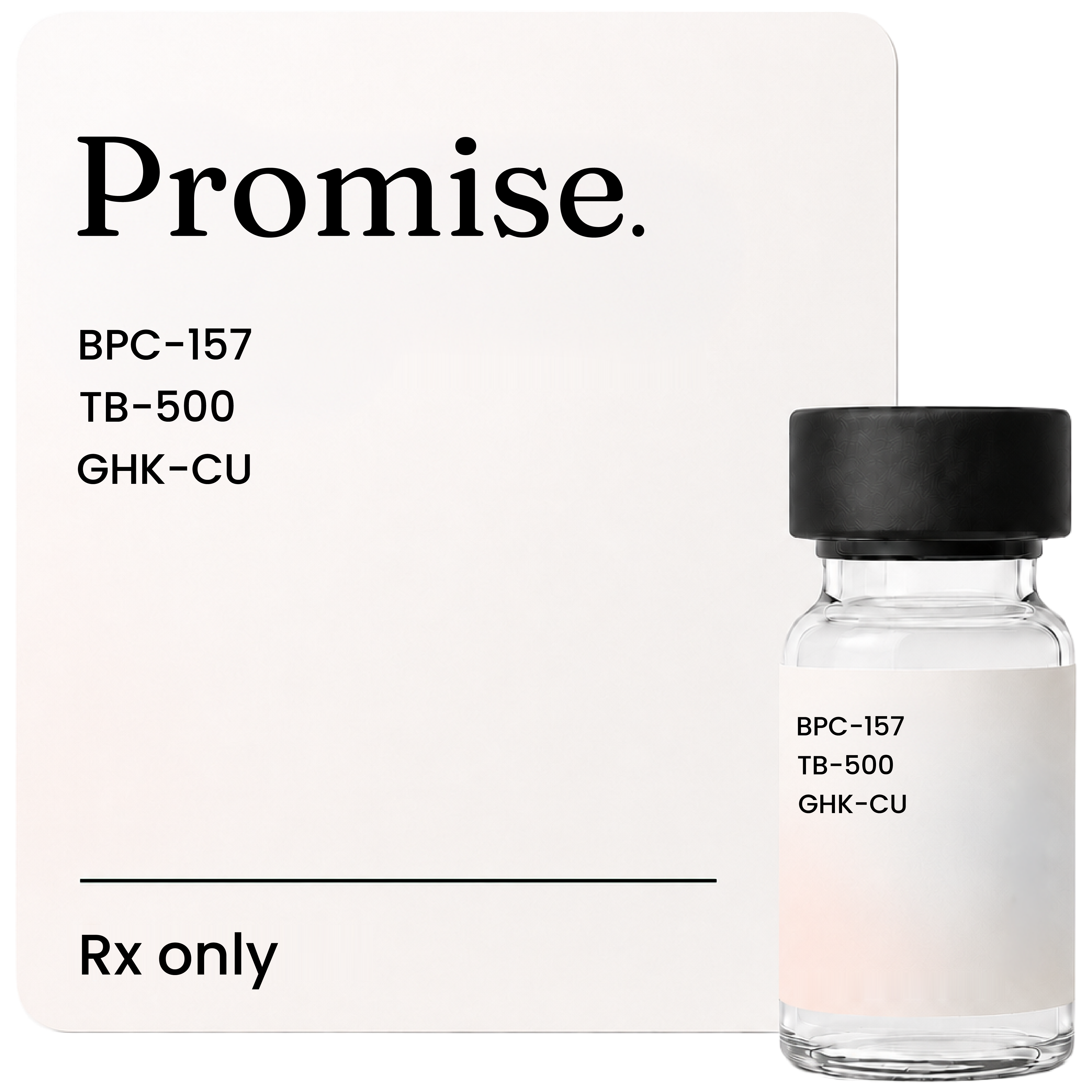
Separate cell and animal tests can't show how three peptides act together in one person. At Promise Peptides (mypromise.com), GLOW (research blend) goes through a prescription route: a licensed clinician looks at each person first and decides. That review doesn't stand in for the missing blend trial.
Wound healing peptides differ: GHK-Cu, BPC-157, and TB-500
Which peptide has the best wound record? The answer depends on the tissue.
BPC-157 has more than thirty animal studies [4][6][8][21]. Its clearest work involves rat tendons, ligaments, and the gut.
TB-500 comes from thymosin beta-4. The parent protein has strong skin, eye, and heart-wound work [9][10][11][R4].
It reached Phase 3 trials. Much of that work concerns cells moving over a wound.
GHK-Cu has the broadest gene work. About 4,000 genes changed in lab tests [14][15][16].
Its best human work used skin creams compared with placebo. Those studies looked at collagen and skin feel.
No study put all three in the same fair test. So you can't rank them with one score.
Each was studied in different tissue and under different rules. A result in an eye isn't a result in your knee.
BPC-157 is best known for tendon and ligament work. TB-500 is best known for skin and eye wound cover.
GHK-Cu is best known for skin collagen and scar repair. Their strengths don't prove a blend.
The BPC-157 TB-500 blend page explains why people pair the first two. It then shows what GHK-Cu might add.
BPC-157 and TB-500 still lack a direct pair test. That keeps the limit clear.